Voluminous Posterior Medial Meniscal Cyst

The human knee, a complex and fascinating joint, is prone to various injuries and conditions that can affect its function and overall health. One such condition is the development of a meniscal cyst, specifically a voluminous posterior medial meniscal cyst. To understand the implications and management of this condition, it’s essential to delve into the anatomy of the knee, the role of the meniscus, and the specifics of meniscal cysts.
Introduction to Knee Anatomy and Meniscal Function
The knee joint is formed by the articulation of the femur (thigh bone) and the tibia (shin bone), with the patella (kneecap) situated at the front. The menisci are two semi-lunar cartilages located between the ends of the bones, one on the medial (inner) aspect and one on the lateral (outer) aspect of the knee. These cartilages play a crucial role in absorbing shock, distributing weight evenly, and facilitating smooth movement of the knee.
Understanding Meniscal Cysts
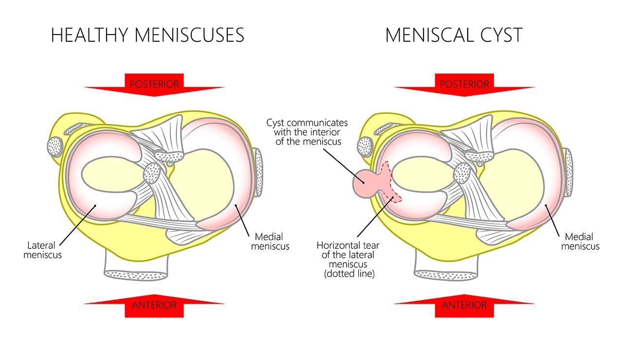
A meniscal cyst is an abnormal fluid-filled structure that can develop in association with a meniscus, most commonly as a result of a meniscal tear. These cysts can form when fluid from the joint (synovial fluid) penetrates through a tear in the meniscus and accumulates, forming a cyst. Meniscal cysts can cause a range of symptoms, including knee pain, swelling, and locking or catching sensations in the knee, depending on their location and size.
Voluminous Posterior Medial Meniscal Cyst: Specifics and Implications
A voluminous posterior medial meniscal cyst is a significant cyst formation located at the posterior (rear) aspect of the medial meniscus. The term “voluminous” indicates that the cyst is particularly large, which can exacerbate symptoms and complicate treatment. The posterior location can pose challenges for diagnosis and treatment, as it may not be as readily apparent on initial examination or standard imaging views.
Symptoms and Diagnosis
Symptoms of a voluminous posterior medial meniscal cyst can include pain on the inner aspect of the knee, which may worsen with activities that involve bending or twisting. A noticeable lump or swelling may be palpable on the inner aspect of the knee, although this can vary depending on the cyst’s exact location and the patient’s body type. Diagnosis typically involves a combination of clinical examination, where a healthcare provider assesses the knee for signs of meniscal injury or cyst formation, and imaging studies such as MRI (Magnetic Resonance Imaging), which can provide detailed images of the knee structures, including meniscal tears and cysts.
Treatment Options
Treatment of a voluminous posterior medial meniscal cyst depends on the severity of symptoms, the presence of underlying meniscal tears, and the patient’s overall health and preferences. Conservative management may involve physical therapy to improve knee function and reduce pain, along with possible corticosteroid injections to decrease inflammation. However, for many patients, surgical intervention is necessary to remove the cyst and address any underlying meniscal tears. Arthroscopic surgery, a minimally invasive procedure where a surgeon uses a small camera and instruments to operate through tiny incisions, is often the preferred method for treating meniscal cysts and tears.
Comparative Analysis of Surgical Approaches
When considering surgical treatment for a voluminous posterior medial meniscal cyst, it’s essential to evaluate the different approaches available. Traditional open surgery involves a larger incision to access the knee joint directly, whereas arthroscopic surgery offers a less invasive alternative with smaller incisions and potentially faster recovery times. Each approach has its advantages and disadvantages, including differences in post-operative pain, rehabilitation periods, and risks of complications.
Arthroscopic Surgery
Arthroscopic surgery is favored for its minimally invasive nature, which can lead to less tissue damage and trauma to the knee. Through small incisions, a surgeon can insert an arthroscope (a thin, flexible tube with a camera and light) and surgical instruments to visualize the knee joint and perform the necessary repairs. This approach allows for precise removal of the cyst and repair of the meniscal tear, promoting healing and reducing the likelihood of future complications.
Open Surgery
In some cases, open surgery might be recommended, especially if the cyst is very large or if there are significant meniscal tears that require more extensive repair. Open surgery provides a more direct access to the knee joint, allowing for a potentially more thorough removal of the cyst and repair of the meniscus. However, this approach is generally associated with a longer recovery period and higher risk of post-operative complications.
Historical Evolution of Meniscal Cyst Treatment
The understanding and treatment of meniscal cysts have undergone significant evolution over the years. Historically, meniscal cysts were often treated with open surgery, which, while effective, carried higher risks of complications and longer recovery periods. The advent of arthroscopic techniques marked a significant advancement, offering a less invasive alternative with improved outcomes. Continuing research and advancements in orthopedic surgery are expected to refine treatment options further, potentially incorporating new technologies and techniques that enhance patient outcomes and minimize recovery times.
Future Trends Projection
Looking ahead, the management of voluminous posterior medial meniscal cysts is likely to incorporate more personalized treatment approaches, leveraging advancements in imaging and diagnostic technologies to tailor interventions to the specific needs of each patient. The integration of regenerative medicine, focusing on the body’s natural healing processes to repair or replace damaged tissues, may also play a role in future treatments. Furthermore, the development of new surgical instruments and techniques, possibly including robotic-assisted surgeries, could offer improved precision and outcomes for patients undergoing surgery for meniscal cysts and tears.
Technical Breakdown: Surgical Procedure
The surgical procedure for removing a voluminous posterior medial meniscal cyst involves several key steps:
Preparation: The patient is prepared for surgery, which includes administering anesthesia and positioning the patient to allow optimal access to the knee.
Arthroscopic Portal Placement: Small incisions are made to insert the arthroscope and surgical instruments. The placement of these portals is critical to ensure adequate visualization and access to the meniscal cyst.
Cyst Identification and Removal: The meniscal cyst is identified, and the surgeon carefully removes it, taking care to avoid damaging surrounding structures.
Meniscal Tear Repair: If a meniscal tear is present, the surgeon will repair it, using techniques such as suture repair or meniscectomy (partial or total removal of the meniscus), depending on the tear’s location and severity.
Postoperative Care: After surgery, the patient begins a rehabilitation program to restore knee function and strength, minimize the risk of complications, and promote healing.
Decision Framework for Treatment
When deciding on the treatment for a voluminous posterior medial meniscal cyst, several factors must be considered:
- Symptom Severity: The degree of pain, swelling, and functional impairment.
- Cyst Size and Location: Larger cysts, especially those in posterior locations, may require more aggressive treatment.
- Presence of Meniscal Tears: The need to address underlying meniscal pathology.
- Patient Health and Preferences: Overall health, age, activity level, and personal preferences regarding treatment options.
- Potential Risks and Benefits: Weighing the advantages and disadvantages of each treatment approach.
Myth vs. Reality: Common Misconceptions about Meniscal Cysts
There are several misconceptions about meniscal cysts, including the belief that they always require surgical intervention or that they are a sign of a more serious underlying condition. In reality, while surgery is often necessary, especially for symptomatic cysts, not all meniscal cysts require immediate surgical treatment. Furthermore, the presence of a meniscal cyst does not inherently indicate a severe underlying condition but rather a response to a meniscal tear or other joint pathology.
Resource Guide: Further Reading and Support
For patients and families seeking more information on meniscal cysts and their treatment, several resources are available:
- Orthopedic Societies: Professional organizations, such as the American Academy of Orthopaedic Surgeons (AAOS), provide patient education materials and resources.
- Peer-Reviewed Journals: Publications like the Journal of Orthopaedic and Sports Physical Therapy and the American Journal of Sports Medicine offer in-depth articles on the latest research and treatment approaches.
- Support Groups: Online forums and local support groups can provide valuable insights and connections with others who have experienced similar conditions.
FAQ Section
What are the symptoms of a voluminous posterior medial meniscal cyst?
+Symptoms can include pain on the inner aspect of the knee, swelling, and a palpable lump. Activities that involve bending or twisting may exacerbate pain.
How is a voluminous posterior medial meniscal cyst diagnosed?
+Diagnosis typically involves a clinical examination and imaging studies, most commonly an MRI, to visualize the cyst and any associated meniscal tears.
What are the treatment options for a voluminous posterior medial meniscal cyst?
+Treatment options include conservative management with physical therapy and corticosteroid injections, as well as surgical intervention, either arthroscopic or open surgery, to remove the cyst and repair any meniscal tears.
What is the recovery time after surgery for a voluminous posterior medial meniscal cyst?
+Recovery time can vary depending on the surgical approach and the extent of the procedure. Generally, arthroscopic surgery offers a faster recovery compared to open surgery, but both require a period of rehabilitation to restore knee function and strength.
Can a voluminous posterior medial meniscal cyst recur after treatment?
+Recurrence is possible, especially if the underlying meniscal tear is not adequately addressed. Proper treatment and rehabilitation are crucial to minimizing the risk of recurrence and ensuring the best possible outcome.
Conclusion
A voluminous posterior medial meniscal cyst presents a unique challenge in the field of orthopedic surgery, requiring a thoughtful and individualized approach to treatment. By understanding the anatomy of the knee, the function of the meniscus, and the specifics of meniscal cysts, patients and healthcare providers can work together to develop effective treatment plans. As research and technology continue to advance, the management of meniscal cysts is likely to evolve, offering improved outcomes and Quality of Life for those affected by this condition.


