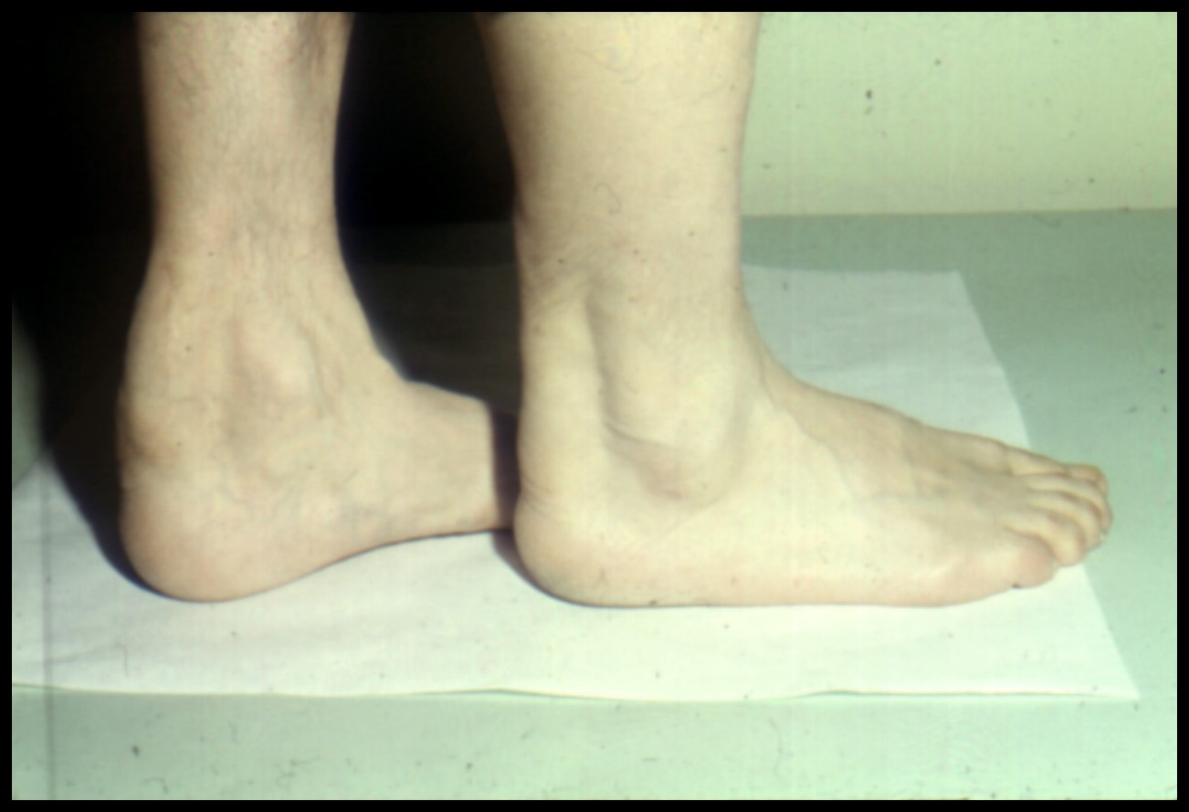
Tendon Xanthomas Images

Tendon xanthomas are characterized by the accumulation of lipid-laden cells, known as foamy histiocytes, within tendons. This condition is often associated with high levels of low-density lipoprotein (LDL) cholesterol, a key component of the lipid profile. Understanding the nature of tendon xanthomas requires an examination of their clinical presentation, pathophysiology, and the challenges in managing these lesions.
Clinical Presentation
Patients with tendon xanthomas typically present with palpable, firm nodules or masses along the course of tendons. These lesions are most commonly found in the Achilles tendons, but they can also occur in the tendons of the hands, feet, and other parts of the body. The presence of these lesions can be asymptomatic, or they may cause discomfort or pain, especially if they are large or if they impinge on surrounding structures.
Pathophysiology
The development of tendon xanthomas is closely linked to abnormalities in lipid metabolism. High levels of LDL cholesterol can lead to the accumulation of lipid within cells of the tendon, leading to the formation of xanthomas. These lesions are composed of lipid-laden macrophages (foamy histiocytes), fibroblasts, and other cellular components. The exact mechanism by which high cholesterol levels lead to the formation of tendon xanthomas is not fully understood, but it is believed to involve the accumulation of lipids in macrophages within the tendon tissue.
Management and Treatment
The management of tendon xanthomas primarily focuses on addressing the underlying lipid disorder. This typically involves the use of statins or other lipid-lowering medications to reduce LDL cholesterol levels. In some cases, surgical excision of the xanthoma may be considered, especially if the lesion is causing significant symptoms or if it is compromising the function of the affected tendon. However, surgery may not always be successful in eliminating the problem, as the underlying metabolic disorder can lead to the formation of new xanthomas.
Images and Diagnosis
Diagnosing tendon xanthomas often involves a combination of clinical examination, imaging studies, and, in some cases, biopsy. Imaging modalities such as ultrasound, magnetic resonance imaging (MRI), and computed tomography (CT) scans can help in identifying the location, size, and extent of the xanthomas. These images can also aid in differentiating tendon xanthomas from other types of tendon lesions or tumors.
It's crucial for clinicians to be aware of the association between tendon xanthomas and lipid disorders. Early recognition and appropriate management of the underlying metabolic condition can help in preventing the progression of these lesions and reducing the risk of associated cardiovascular diseases.
Impact on Quality of Life
While tendon xanthomas themselves may not always cause significant symptoms, their presence can indicate an increased risk of cardiovascular events due to the associated high levels of LDL cholesterol. Therefore, managing these lesions not only involves addressing the local tendon pathology but also entails a comprehensive approach to reducing cardiovascular risk factors. This can have a profound impact on the quality of life for patients, as it may require significant lifestyle changes and long-term adherence to medication regimens.
Future Directions
Research into the pathogenesis of tendon xanthomas and their relationship with lipid metabolism may uncover new therapeutic targets. For instance, understanding the molecular mechanisms by which lipid accumulation leads to xanthoma formation could lead to the development of novel treatments that specifically address this process. Additionally, advancements in imaging technologies could improve the diagnosis and monitoring of these lesions, potentially allowing for earlier intervention and better outcomes.
What is the primary treatment for tendon xanthomas?
+The primary treatment for tendon xanthomas involves addressing the underlying lipid disorder, typically through the use of lipid-lowering medications such as statins.
Are tendon xanthomas painful?
+Tendon xanthomas can be asymptomatic, but they may also cause discomfort or pain, especially if they are large or impinge on surrounding structures.
Can tendon xanthomas be removed surgically?
+In conclusion, tendon xanthomas are lesions that occur within tendons due to the accumulation of lipid-laden cells, often in association with high levels of LDL cholesterol. Their management involves a multidisciplinary approach, focusing on the treatment of the underlying lipid disorder, and in some cases, surgical intervention for symptom relief. Ongoing research into the pathophysiology of these lesions may lead to the development of more targeted therapies, improving outcomes for patients with tendon xanthomas.