Digestive System 3D Pictures

The digestive system is a complex and fascinating process that involves the breakdown and absorption of nutrients from the food we eat. To better understand this process, let’s take a closer look at the different parts of the digestive system and how they work together.
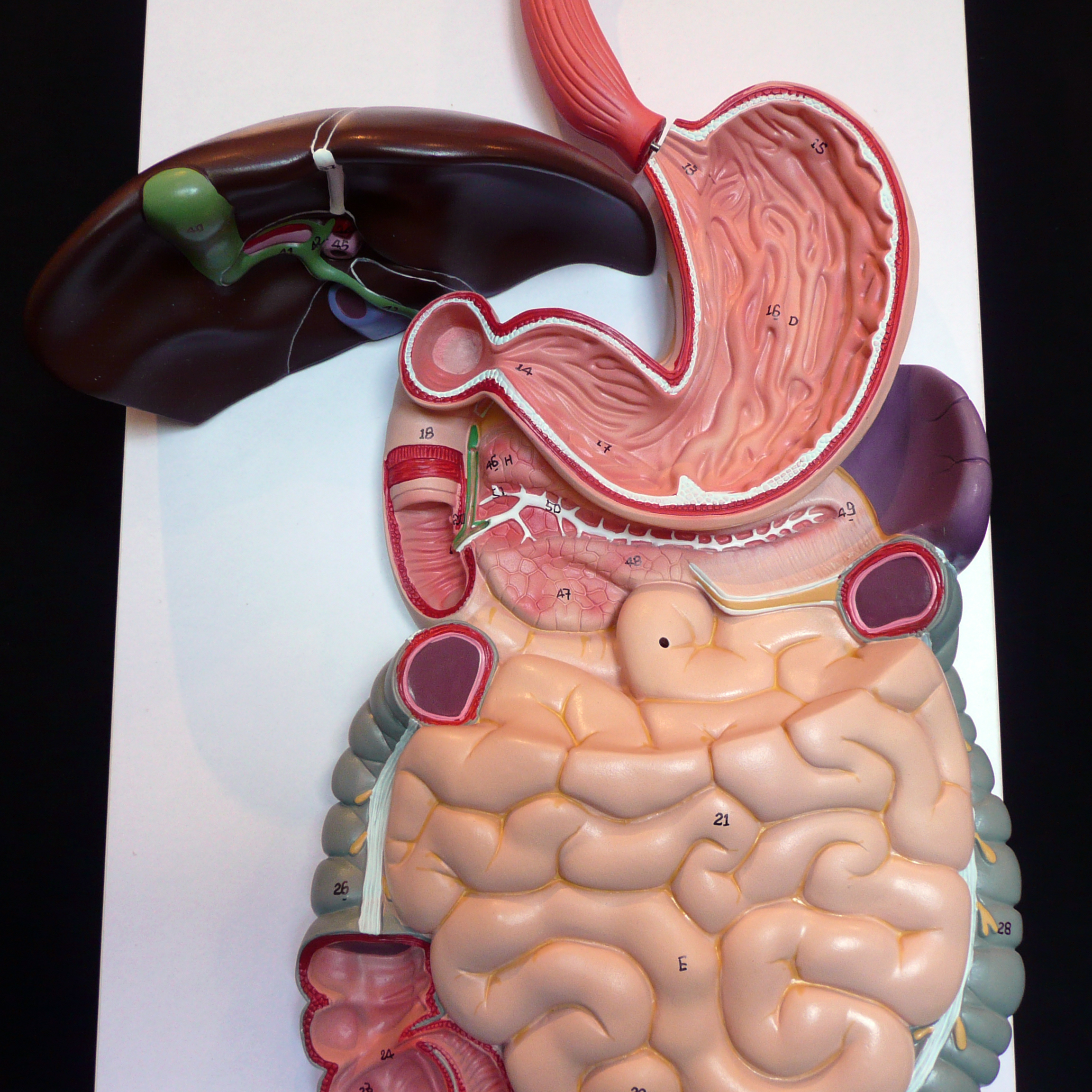
One way to visualize the digestive system is through 3D pictures and models. These can provide a detailed and interactive representation of the different organs and structures involved in digestion, from the mouth and esophagus to the stomach, small intestine, and large intestine.
For instance, a 3D model of the mouth can show how the teeth chew food into smaller pieces, while the tongue mixes this food with saliva that contains enzymes to break down carbohydrates. The esophagus, which is often depicted in 3D as a muscular tube, then propels this food mixture into the stomach through a process known as peristalsis.
In the stomach, 3D pictures can illustrate the lining of the stomach, which secretes mucus to protect itself from the acidic digestive juices, as well as the muscles that churn and mix the food with these juices. The stomach’s role in breaking down proteins and fats into smaller molecules can be visually represented through 3D animations of enzymatic reactions.
Moving further down the digestive tract, the small intestine is where most of our nutrient absorption takes place. 3D models of the small intestine can show its extensive surface area, lined with finger-like projections called villi, which increase the area for absorption. The pancreas and liver also play critical roles in digestion by secreting digestive enzymes and bile, respectively, into the small intestine, and these processes can be vividly depicted through 3D visualizations.
Finally, the large intestine, or colon, absorbs water and electrolytes, and its 3D representations can demonstrate how it compacts the remaining waste material and eliminates it from the body. Understanding the digestive system through 3D pictures not only aids in comprehension but also fosters appreciation for the intricate and highly efficient process that sustains us.
How 3D Pictures Enhance Learning
- Interactive Engagement: 3D pictures encourage active learning by allowing students to interact with the models. This interaction can stimulate curiosity and motivate learners to explore the subject matter in more depth.
- Visual Clarity: Complex anatomical relationships and physiological processes can be difficult to comprehend through textual descriptions alone. 3D models provide a clear visual representation, making it easier for learners to understand spatial relationships and functional dynamics.
- Personalized Learning Experience: With 3D models, educators can tailor the learning experience to the needs and abilities of individual students. For example, students can focus on specific parts of the digestive system that they find challenging, repeating the interactive lessons as many times as needed.
- Accessibility: Digital 3D models can be easily shared and accessed by students with disabilities, improving inclusivity in education. They can also be used in remote learning environments, expanding educational opportunities.
Creating Effective 3D Learning Materials for the Digestive System
- Define Learning Objectives: Clearly outline what learners should understand about the digestive system after interacting with the 3D models.
- Design Interactive Elements: Incorporate features that allow learners to manipulate the 3D models, such as zooming, rotating, and dissecting to explore different layers and structures.
- Integrate Realistic Animations: Use animations to demonstrate dynamic processes like peristalsis, digestion, and absorption, making the learning experience more engaging and effective.
- Provide Contextual Information: Offer accompanying text, audio, or video explanations that provide additional context and insights into the structures and processes being visualized.
- Evaluate and Refine: Conduct usability tests and gather feedback from learners and educators to identify areas for improvement and refine the 3D learning materials accordingly.
The Future of 3D Visualization in Education
As technology continues to advance, we can expect even more sophisticated and interactive 3D visualizations to emerge. Virtual reality (VR) and augmented reality (AR) are already being explored for their potential to revolutionize educational experiences, including the study of the digestive system. These technologies have the potential to transport learners into the human body, where they can explore the digestive system in a fully immersive and interactive environment.
What are the benefits of using 3D pictures to learn about the digestive system?
+The benefits include enhanced understanding through visual and interactive engagement, improved retention of complex information, and the ability to explore the digestive system in a detailed and personalized manner.
How can 3D models be used in educational settings to teach about the digestive system?
+3D models can be integrated into lesson plans as interactive tools for students to explore the digestive system. They can be used to demonstrate processes, illustrate structures, and facilitate discussions and quizzes to assess understanding.
What role can virtual and augmented reality play in the future of learning about the human digestive system?
+Virtual and augmented reality technologies have the potential to provide fully immersive learning experiences. They can simulate the journey of food through the digestive system, allowing learners to explore and understand the process in unprecedented detail and interactivity.
The integration of 3D pictures and models, along with emerging technologies like VR and AR, marks a significant shift in how we approach learning about complex biological systems like the digestive system. By leveraging these tools, we can create learning experiences that are not only more engaging but also more effective, paving the way for a deeper understanding and appreciation of human biology.


