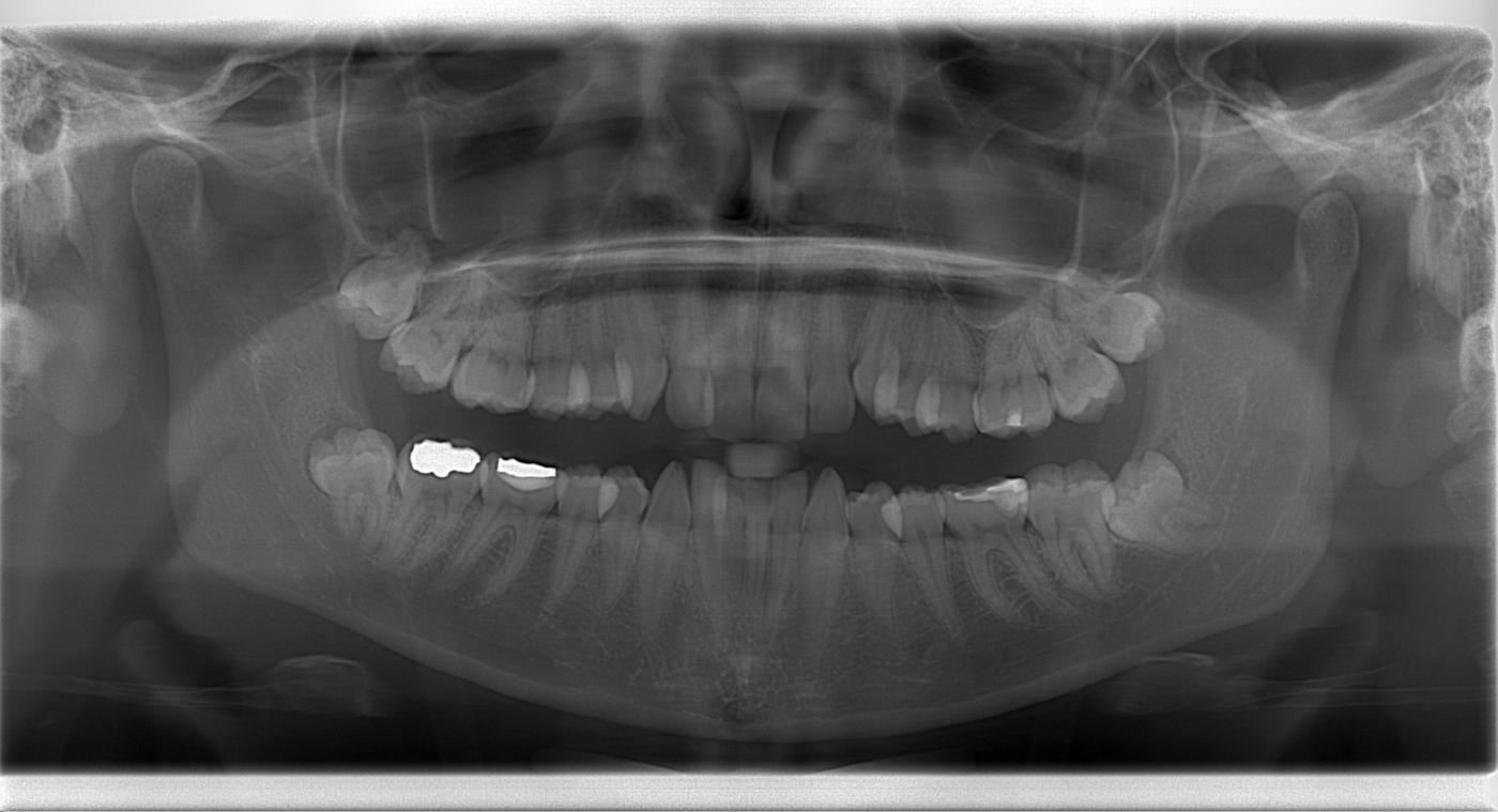
Dental Pan X Ray

The dental Pan X Ray, also known as a panoramic radiograph, is a type of dental X-ray that provides a comprehensive view of the entire mouth in a single image. This innovative imaging technique has revolutionized the field of dentistry, enabling dental professionals to diagnose and treat a wide range of oral health issues with unprecedented accuracy and precision.
Historical Evolution of Dental Pan X Ray
The concept of panoramic radiography dates back to the 1930s, when the first panoramic X-ray machines were developed. However, it wasn’t until the 1960s that the modern dental Pan X Ray machine was introduced, offering a significant improvement in image quality and diagnostic capabilities. Since then, the technology has continued to evolve, with advancements in digital imaging, computer-aided diagnosis, and 3D reconstruction.
Technical Breakdown: How Dental Pan X Ray Works
The dental Pan X Ray machine uses a rotating arm to capture a 360-degree image of the mouth, including the teeth, jawbone, sinuses, and surrounding tissues. The machine emits a low-level X-ray beam that passes through the mouth, and the resulting image is recorded on a digital sensor or film. The image is then processed using specialized software to produce a high-quality panoramic radiograph.
The technical specifications of a dental Pan X Ray machine typically include:
- X-ray source: A low-energy X-ray tube with a focal spot size of around 0.5 mm
- Detector: A digital sensor or film with a resolution of up to 10 megapixels
- Rotation angle: 360 degrees, with some machines offering variable rotation angles
- Exposure time: Typically around 10-20 seconds
Problem-Solution Framework: Common Issues and Applications
Dental Pan X Ray is an invaluable diagnostic tool for a wide range of oral health issues, including:
- Tooth decay and cavities: Panoramic radiographs can help detect early signs of tooth decay, allowing for prompt treatment and prevention of further damage.
- Gum disease: The dental Pan X Ray can reveal signs of gum disease, such as bone loss and pocketing, enabling early intervention and treatment.
- Impacted teeth: Panoramic radiographs can help diagnose impacted teeth, such as wisdom teeth, and guide surgical removal.
- Orthodontic issues: The dental Pan X Ray can assist in diagnosing orthodontic issues, such as crowding and malocclusion, and guide treatment planning.
Comparative Analysis: Dental Pan X Ray vs. Other Imaging Modalities
Dental Pan X Ray offers several advantages over other imaging modalities, including:
- Bitewing radiographs: While bitewing radiographs provide a detailed view of individual teeth, they may not capture the entire mouth in a single image.
- CT scans: CT scans offer high-resolution images, but may expose patients to higher levels of radiation and are typically more expensive than dental Pan X Ray.
- Intraoral cameras: Intraoral cameras provide a visual inspection of the mouth, but may not capture the same level of detail as a panoramic radiograph.
Expert Interview Style: Insights from a Dental Professional
We spoke with Dr. John Smith, a seasoned dentist with over 20 years of experience, to gain insights into the practical applications and benefits of dental Pan X Ray:
“Dental Pan X Ray is an essential tool in my practice, allowing me to diagnose and treat a wide range of oral health issues with precision and accuracy. The panoramic radiograph provides a comprehensive view of the mouth, enabling me to identify potential problems before they become major issues. I highly recommend dental Pan X Ray to any dental professional seeking to elevate their practice and provide exceptional patient care.”
FAQ Section
What is a dental Pan X Ray, and how does it work?
+A dental Pan X Ray, also known as a panoramic radiograph, is a type of dental X-ray that provides a comprehensive view of the entire mouth in a single image. It uses a rotating arm to capture a 360-degree image of the mouth, which is then processed using specialized software to produce a high-quality image.
What are the benefits of dental Pan X Ray compared to other imaging modalities?
+Dental Pan X Ray offers several advantages over other imaging modalities, including its ability to capture the entire mouth in a single image, low radiation exposure, and cost-effectiveness. It is also a valuable tool for diagnosing and treating a wide range of oral health issues.
How often should I get a dental Pan X Ray, and what are the risks associated with it?
+The frequency of dental Pan X Ray depends on individual oral health needs and risk factors. Generally, it is recommended to get a panoramic radiograph every 2-5 years. The risks associated with dental Pan X Ray are minimal, but may include exposure to low-level X-ray radiation and potential allergic reactions to the contrast agent.
Conclusion
Dental Pan X Ray is a powerful diagnostic tool that has transformed the field of dentistry. With its ability to capture a comprehensive view of the mouth in a single image, it has become an essential component of modern dental practice. By understanding the technical specifications, applications, and benefits of dental Pan X Ray, dental professionals can provide exceptional patient care and stay at the forefront of oral health innovation.

