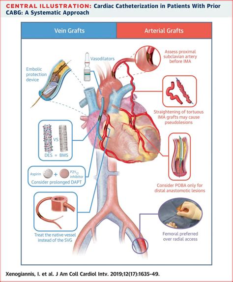
Cardiac Cath Diagram Explained

The cardiac catheterization diagram, often abbreviated as a cardiac cath diagram, is a crucial visual tool used in the field of cardiology. This diagram plays a pivotal role in understanding the procedure of cardiac catheterization, which involves the insertion of a catheter (a thin, flexible tube) into the arteries or veins in the leg or arm and guiding it to the heart. The purpose of this procedure is to diagnose and potentially treat various heart conditions. Let’s delve into the components and interpretations of a cardiac cath diagram, understanding its significance in both diagnostic and interventional cardiology.
Introduction to Cardiac Catheterization
Cardiac catheterization is a procedure that allows cardiologists to visualize the heart’s chambers and vessels. This is achieved by injecting a contrast dye into the catheter, which then becomes visible on an X-ray. The procedure can help diagnose coronary artery disease, measure blood pressure within the heart, assess the pumping function of the heart, and identify structural problems such as septal defects.
Components of a Cardiac Cath Diagram
A typical cardiac cath diagram includes several key components, each providing critical information about the heart’s structure and function:
Arterial and Venous Access: The diagram usually starts with the point of access, where the catheter is inserted. This is typically in the femoral artery in the groin or the radial artery in the wrist for arterial access, and in a vein for venous access.
Catheter Pathway: The pathway the catheter takes through the blood vessels to reach the heart. This includes navigating through the aorta to reach the coronary arteries or through the superior and inferior vena cava to reach the right side of the heart.
Heart Chambers: The diagram will show the four chambers of the heart: the right atrium, right ventricle, left atrium, and left ventricle. Each chamber can be assessed for size, wall motion, and pressure.
Valves: The heart has four valves (tricuspid, pulmonary, mitral, and aortic), which are crucial for ensuring blood flows in one direction. The diagram will show the location and function of these valves.
Coronary Arteries: For procedures focusing on coronary artery disease, the diagram will detail the coronary arteries’ anatomy, including any blockages or narrowing.
Contrast Dye Injection: The point at which contrast dye is injected to visualize the heart’s structures on an X-ray.
Interpreting a Cardiac Cath Diagram
Interpreting a cardiac cath diagram requires a thorough understanding of cardiac anatomy and the principles of cardiac catheterization. Key areas of focus include:
Pressure Recordings: The diagram may include pressure tracings from different chambers of the heart, which can indicate abnormalities such as stenotic valves or ventricular dysfunction.
Angiographic Images: These are the X-ray images taken after contrast dye injection, showing the inside of the heart’s chambers and vessels. They can reveal structural abnormalities, vessel blockages, or issues with heart function.
Blood Flow and Oxygenation: Information about blood flow through different parts of the heart and lungs, as well as oxygen saturation levels, can be critical for diagnosing certain conditions.
Applications in Diagnostic and Interventional Cardiology
Cardiac cath diagrams are not only essential for diagnostic purposes but also play a crucial role in interventional cardiology, guiding procedures such as:
Angioplasty and Stenting: Using the diagram to navigate to blocked coronary arteries and perform angioplasty (expanding the artery with a balloon) followed by stenting (placing a wire mesh tube to keep the artery open).
Valvuloplasty: Similar to angioplasty but used for narrowed heart valves.
Closure of Septal Defects: Using the catheter to deliver a device that closes holes between the heart’s chambers.
Conclusion
The cardiac cath diagram is a fundamental tool in cardiology, serving as a visual representation of the cardiac catheterization procedure. It aids in the diagnosis of various cardiac conditions and guides interventional procedures to treat these conditions. Understanding the components and interpretation of a cardiac cath diagram is crucial for healthcare professionals involved in cardiology, as it directly impacts patient care and outcomes.
FAQ Section
What is the primary purpose of a cardiac catheterization diagram?
+The primary purpose of a cardiac catheterization diagram is to visualize the heart’s chambers and vessels, aiding in the diagnosis and treatment of heart conditions through procedures like cardiac catheterization.
What are the key components of a cardiac cath diagram?
+A cardiac cath diagram includes components such as arterial and venous access points, the catheter pathway, heart chambers, valves, coronary arteries, and the point of contrast dye injection.
How does a cardiac cath diagram aid in interventional cardiology?
+A cardiac cath diagram guides interventional procedures by providing detailed anatomical information, allowing for precise navigation to treat conditions such as coronary artery blockages, valve issues, and septal defects.
What is the significance of interpreting pressure recordings and angiographic images in a cardiac cath diagram?
+Interpreting pressure recordings and angiographic images is crucial for diagnosing abnormalities such as valve stenosis, ventricular dysfunction, and coronary artery disease, guiding appropriate therapeutic interventions.
Can cardiac catheterization diagnose all types of heart conditions?
+While cardiac catheterization is a powerful diagnostic tool, it is primarily focused on conditions related to the coronary arteries, heart chambers, and valves. Other conditions may require additional diagnostic tests.
