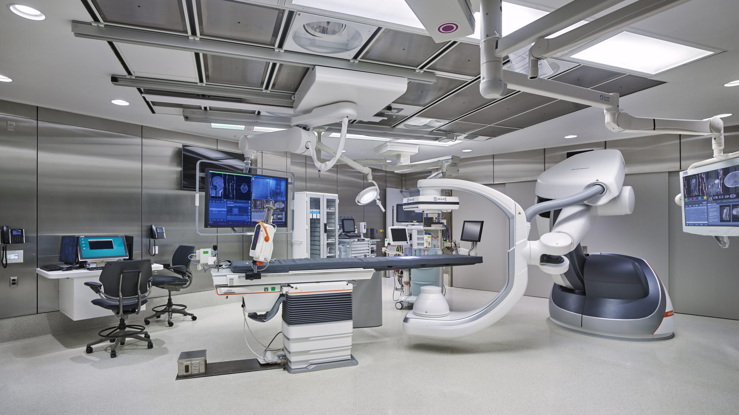
Cardiac Cath Diagram

The cardiac catheterization procedure, commonly referred to as a cardiac cath, is a vital diagnostic tool used in cardiology to visualize the heart’s chambers and vessels. This process involves inserting a catheter, a thin, flexible tube, through an artery in the leg or arm and guiding it to the heart. Once in place, the catheter can be used for various purposes, including injecting dye into the heart’s vessels to create detailed images of its structure and function.
Understanding the Cardiac Catheterization Process
Preparation: Before the procedure, the patient is prepared by having the area where the catheter will be inserted cleaned and numbed with local anesthesia. The patient may also be given sedation to help relax.
Insertion: The doctor then inserts the catheter through a small incision in the skin and into the artery. Using real-time X-ray images (fluoroscopy), the catheter is guided through the arterial system to the heart.
Dye Injection: Once the catheter is in the desired position, a special dye (contrast agent) is injected through it. This dye makes the heart’s chambers and vessels visible on X-ray images, allowing the doctor to see any abnormalities, such as blockages in the coronary arteries.
Monitoring and Diagnosis: The procedure is monitored closely, and images are recorded for later review. Based on the findings, the doctor can diagnose conditions like coronary artery disease, heart valve problems, or congenital heart defects.
Interventional Procedures: If necessary, the cardiac cath can also be used to perform certain treatments, such as angioplasty (to open narrowed arteries) or stenting (to keep arteries open).
The Cardiac Cath Diagram Explained
A typical cardiac cath diagram illustrates the pathway of the catheter from the insertion point in the leg or arm, through the arterial system, and into the heart. Key components usually depicted include:
- Arterial Access: The point where the catheter enters the body, typically through the femoral artery in the groin or the radial artery in the wrist.
- Aorta: The main artery that arises from the heart’s left ventricle, branching into smaller arteries that supply blood to the body.
- Coronary Arteries: The arteries that branch off the aorta to supply blood directly to the heart muscle itself. These are often the focus of cardiac catheterization procedures.
- Heart Chambers: The procedure can visualize the four chambers of the heart (right and left atria, and right and left ventricles) to assess their function and structure.
- Valves: The heart has four valves (mitral, tricuspid, pulmonary, and aortic) that ensure blood flows in one direction. The cardiac cath can help evaluate valve function.
Advanced Techniques and Technologies
The field of cardiac catheterization continues to evolve, with advancements in imaging technologies, catheter design, and the development of minimally invasive procedures. Techniques such as fractional flow reserve (FFR) measurements to assess the severity of blockages and intravascular ultrasound (IVUS) for detailed imaging of the arteries’ inner walls are becoming more prevalent.
Future Directions
As medical technology advances, the cardiac catheterization procedure is expected to become even more sophisticated. Emerging trends include the use of robotics in catheter navigation, enhanced imaging modalities that provide real-time detailed views of the heart’s anatomy, and more effective and safer treatments for heart conditions.
Conclusion
The cardiac cath diagram represents a crucial tool in the diagnosis and treatment of heart diseases. By understanding the components and process involved in cardiac catheterization, patients and healthcare professionals can better appreciate the complexities and benefits of this procedure. As advancements continue, the role of cardiac catheterization in managing cardiac health is likely to expand, offering new hope for individuals with heart conditions.
What is the purpose of injecting dye during a cardiac catheterization?
+The dye, or contrast agent, is used to make the heart’s chambers and blood vessels visible on X-ray images, allowing doctors to identify any blockages, abnormalities, or other issues within the heart.
Is cardiac catheterization a risky procedure?
+Like any invasive medical procedure, cardiac catheterization carries risks, including bleeding, infection, and reaction to the contrast dye. However, for most patients, the benefits of the procedure in terms of diagnosis and treatment of heart conditions far outweigh the risks.
How long does a cardiac catheterization procedure typically take?
+The duration of the procedure can vary depending on what is being done. A diagnostic cardiac catheterization might take about 30 minutes to an hour, while more complex procedures, like angioplasty and stenting, can take longer, up to several hours.


