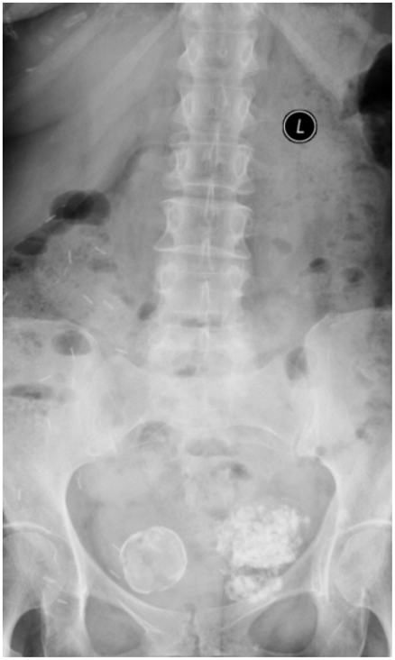
Are Calcified Uterine Fibroids Dangerous

Uterine fibroids are a common health issue affecting many women, particularly during their reproductive years. These growths, which can range in size from small to quite large, develop in or around the uterus and are usually benign. However, when uterine fibroids undergo calcification, a process where calcium deposits form within the fibroid, it can raise concerns about potential health implications. Understanding the nature of calcified uterine fibroids and their potential risks is essential for managing symptoms and maintaining overall health.
What are Uterine Fibroids?
Before delving into the specifics of calcified uterine fibroids, it’s helpful to understand what uterine fibroids are. Uterine fibroids, also known as leiomyomas, are noncancerous growths that appear in or on the uterus. They can vary significantly in size, from small, pea-sized growths that are barely detectable to large masses that can distort the shape of the uterus. The exact cause of uterine fibroids is not well understood, but several factors, including hormonal influences, genetic predisposition, and environmental factors, are believed to contribute to their development.
What is Calcification in Uterine Fibroids?
Calcification refers to the accumulation of calcium salts in a body tissue. This process can occur in various parts of the body and is often seen in the context of injury, aging, or certain disease processes. In the case of uterine fibroids, calcification typically occurs as the fibroid ages. It’s a degenerative process where the fibroid tissue undergoes changes, leading to the deposition of calcium. Calcified uterine fibroids can be detected through imaging studies such as ultrasound, CT scans, or MRI, which can identify the calcium deposits within the fibroid.
Symptoms and Risks Associated with Calcified Uterine Fibroids
While most uterine fibroids, including those that are calcified, are benign and do not lead to serious health issues, their presence can be associated with several symptoms and potential risks. Symptoms may include:
- Pelvic Pressure or Pain: Large calcified fibroids can exert pressure on surrounding organs, leading to discomfort or pain.
- Heavy or Prolonged Menstrual Bleeding: Although less common with calcified fibroids, significant bleeding can still occur, especially if the fibroid is located in a position that affects uterine blood flow.
- Urinary Frequency: Pressure on the bladder can lead to a need to urinate more frequently.
- Constipation: Large fibroids can also press on the bowel, leading to constipation.
Regarding risks, the primary concern with calcified uterine fibroids is not typically their potential to become malignant but rather the symptoms and complications they can cause. However, in rare instances, a condition known as “red degeneration” can occur, especially during pregnancy, leading to severe pain. Additionally, very large fibroids, even if calcified, can potentially lead to complications such as infertility or pregnancy complications due to their size and the pressure they exert on the uterus and surrounding structures.
Management and Treatment Options
The management of calcified uterine fibroids depends on several factors, including the size of the fibroid, the severity of symptoms, and the woman’s overall health and reproductive plans. For many women, especially those without significant symptoms, a “wait and watch” approach with regular check-ups may be recommended. This approach involves monitoring the fibroid’s size and the woman’s symptoms over time.
For those experiencing significant symptoms, several treatment options are available, including:
- Hysterectomy: Surgical removal of the uterus. This is a definitive treatment but is typically considered for severe cases.
- Myomectomy: Surgical removal of the fibroids while leaving the uterus in place. This can be an option for women wishing to preserve their fertility.
- Uterine Artery Embolization (UAE): A minimally invasive procedure that cuts off the blood supply to the fibroid, causing it to shrink.
- Magnetic Resonance-guided Focused Ultrasound (MRgFUS): A non-invasive procedure that uses high-frequency sound waves to heat and destroy the fibroid tissue.
Conclusion
Calcified uterine fibroids, while not typically dangerous in the sense of being cancerous, can still cause significant symptoms and complications for some women. Understanding the nature of these growths and being aware of the available management and treatment options can help in making informed decisions about health care. For women experiencing symptoms or concerns related to uterine fibroids, consulting with a healthcare provider is essential to discuss the best course of action based on individual circumstances.
What causes uterine fibroids to become calcified?
+Calcification in uterine fibroids typically occurs as the fibroid ages, involving a degenerative process where calcium deposits form within the fibroid tissue. The exact triggers for calcification are not fully understood but are believed to be related to the natural aging process of the fibroid and possibly influenced by hormonal changes.
Can calcified uterine fibroids affect fertility?
+Large calcified fibroids, especially those that are located within the uterine cavity or significantly distort the uterine shape, can potentially impact fertility. However, this effect can vary greatly from one woman to another, depending on the size, location, and number of fibroids. Consulting a healthcare provider or a fertility specialist is recommended for personalized advice.
How are calcified uterine fibroids diagnosed?
+Diagnosis of calcified uterine fibroids often involves imaging studies. Ultrasound is commonly used due to its availability and effectiveness in visualizing the uterus and detecting fibroids. However, CT scans or MRI may be employed for more detailed evaluation, especially when assessing the size and location of the fibroid or when planning for surgical intervention.


