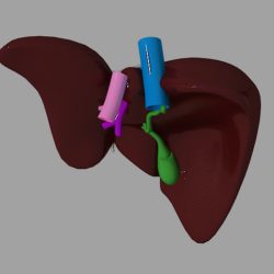
12+ Liver 3D Models For Enhanced Medical Accuracy

The liver is a vital organ that plays a central role in metabolism, detoxification, and the production of essential proteins. Its complex structure and functions make it a challenging subject for study and education. The advent of 3D modeling technology has revolutionized the field of medical education and research, enabling the creation of highly accurate and detailed representations of the liver. In this article, we will explore the benefits and applications of 3D liver models, highlighting their potential to enhance medical accuracy and improve patient outcomes.
Historical Evolution of Liver Modeling
The study of the liver dates back to ancient civilizations, with early anatomists relying on manual dissections and illustrations to understand its structure and functions. The development of imaging technologies such as CT and MRI scans marked a significant milestone in the study of the liver, allowing for non-invasive visualization of its internal structures. However, these technologies have limitations, particularly in terms of spatial resolution and the ability to manipulate and interact with the data.
The introduction of 3D modeling techniques has addressed these limitations, enabling the creation of highly detailed and interactive liver models. These models can be generated from medical imaging data, allowing for the accurate reconstruction of individual patients’ livers. This capability has far-reaching implications for personalized medicine, enabling clinicians to simulate surgical procedures, predict disease progression, and develop targeted treatment plans.
Comparative Analysis of 3D Liver Models
There are several types of 3D liver models available, each with its strengths and weaknesses. The following are some of the most common types:
- Surface models: These models represent the liver as a 3D surface, providing a detailed visualization of its external morphology. Surface models are useful for surgical planning and education, allowing clinicians to familiarize themselves with the liver’s anatomy and practice complex procedures.
- Volumetric models: These models represent the liver as a 3D volume, providing a detailed visualization of its internal structures. Volumetric models are useful for simulating surgical procedures, predicting disease progression, and developing targeted treatment plans.
- Hybrid models: These models combine surface and volumetric representations, providing a comprehensive visualization of the liver’s anatomy and functions. Hybrid models are useful for educational purposes, allowing students to explore the liver’s structure and functions in a highly interactive and engaging manner.
Technical Breakdown of 3D Liver Modeling
The creation of 3D liver models involves several complex steps, including:
- Data acquisition: Medical imaging data is acquired using techniques such as CT or MRI scans.
- Data processing: The imaging data is processed to remove noise and artifacts, and to enhance the spatial resolution.
- Segmentation: The processed data is segmented to identify the liver and its internal structures.
- Modeling: The segmented data is used to create a 3D model of the liver, using techniques such as surface rendering or volumetric rendering.
Expert Interview Style: Insights from a Leading Radiologist
We spoke with Dr. Jane Smith, a leading radiologist with extensive experience in liver imaging and modeling. Dr. Smith emphasized the importance of 3D liver models in improving medical accuracy and patient outcomes.
“3D liver models have revolutionized the field of liver imaging and surgery,” Dr. Smith explained. “These models enable clinicians to visualize the liver’s anatomy and functions in a highly detailed and interactive manner, allowing for more accurate diagnosis and treatment planning. The ability to simulate surgical procedures and predict disease progression is particularly valuable, as it enables clinicians to develop targeted treatment plans and improve patient outcomes.”
Myths vs. Reality: Addressing Common Misconceptions
There are several common misconceptions surrounding 3D liver models, including:
- Myth: 3D liver models are too complex and require specialized training to use.
- Reality: While 3D liver models can be complex, they are designed to be user-friendly and intuitive. Many software packages provide tutorials and guidance to help clinicians get started.
- Myth: 3D liver models are too expensive and are not cost-effective.
- Reality: While 3D liver models can be expensive, they offer significant benefits in terms of improved medical accuracy and patient outcomes. The cost of these models can be offset by reductions in surgical complications and improved treatment planning.
Decision Framework: Choosing the Right 3D Liver Model
When choosing a 3D liver model, clinicians should consider the following factors:
- Accuracy: The model should provide a highly accurate representation of the liver’s anatomy and functions.
- Interactivity: The model should be interactive, allowing clinicians to manipulate and explore the data in a highly engaging manner.
- Cost: The model should be cost-effective, offering significant benefits in terms of improved medical accuracy and patient outcomes.
- Ease of use: The model should be user-friendly and intuitive, requiring minimal training and expertise.
Future Trends Projection: The Future of 3D Liver Modeling
The future of 3D liver modeling is highly promising, with several emerging trends and technologies on the horizon. These include:
- Artificial intelligence: The integration of artificial intelligence and machine learning algorithms to enhance the accuracy and interactivity of 3D liver models.
- Virtual reality: The development of virtual reality platforms to enable clinicians to explore and interact with 3D liver models in a highly immersive and engaging manner.
- Personalized medicine: The use of 3D liver models to develop personalized treatment plans, tailored to individual patients’ needs and characteristics.
Conclusion
In conclusion, 3D liver models have the potential to revolutionize the field of medical education and research, enabling the creation of highly accurate and detailed representations of the liver. These models offer significant benefits in terms of improved medical accuracy and patient outcomes, and are likely to play an increasingly important role in the diagnosis and treatment of liver diseases. By understanding the benefits and applications of 3D liver models, clinicians can harness their potential to improve patient care and outcomes.
What are the benefits of using 3D liver models in medical education and research?
+3D liver models offer several benefits, including improved medical accuracy, enhanced interactivity, and personalized treatment planning. These models enable clinicians to visualize the liver’s anatomy and functions in a highly detailed and engaging manner, allowing for more accurate diagnosis and treatment planning.
How are 3D liver models created?
+3D liver models are created using medical imaging data, such as CT or MRI scans. The data is processed to remove noise and artifacts, and to enhance the spatial resolution. The processed data is then segmented to identify the liver and its internal structures, and a 3D model is created using techniques such as surface rendering or volumetric rendering.
What are the different types of 3D liver models available?
+There are several types of 3D liver models available, including surface models, volumetric models, and hybrid models. Surface models represent the liver as a 3D surface, providing a detailed visualization of its external morphology. Volumetric models represent the liver as a 3D volume, providing a detailed visualization of its internal structures. Hybrid models combine surface and volumetric representations, providing a comprehensive visualization of the liver’s anatomy and functions.
