12 Cervical Polyp Images For Accurate Diagnosis

Cervical polyps are growths that occur on the cervix, which is the lower part of the uterus in the human female reproductive system. These polyps are usually benign (non-cancerous) but can cause symptoms such as abnormal vaginal bleeding, particularly after intercourse, menstrual periods, or douching. For an accurate diagnosis, healthcare providers often rely on a combination of clinical history, physical examination, and diagnostic imaging. Here, we’ll explore 12 cervical polyp images that can aid in understanding and diagnosing these growths, emphasizing the importance of visual recognition in medical practice.
1. Cervical Polyp on Colposcopy
- Image Description: A colposcopy image showing a small, rounded polyp on the cervix. The polyp appears as a distinct, protruding lesion.
- Diagnosis: This image highlights the use of colposcopy, a procedure that uses a special microscope (colposcope) to examine the cervix, vagina, and vulva for signs of disease. The image here demonstrates how colposcopy can help identify cervical polyps.
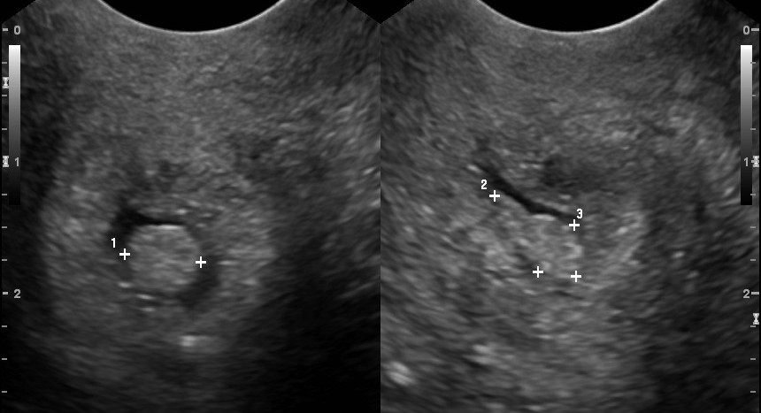
2. Uterus with Cervical Polyp on Ultrasound
- Image Description: An ultrasound image of the uterus showing a cervical polyp. The polyp is visible as a small, hypoechoic (darker) area attached to the cervical canal.
- Diagnosis: Ultrasound imaging is crucial for assessing the size and location of cervical polyps, as well as for evaluating the uterine cavity and surrounding structures.
3. Endocervical Polyp on Hysteroscopy
- Image Description: A hysteroscopic image showing an endocervical polyp within the cervical canal. The polyp appears as a small, pedunculated mass.
- Diagnosis: Hysteroscopy, which involves the insertion of a telescope (hysteroscope) through the cervix to view the inside of the uterus, is particularly useful for diagnosing and potentially treating endocervical polyps.
4. Cervical Polyp on MRI
- Image Description: An MRI image of the pelvis showing a cervical polyp as a small, well-defined mass on the cervix.
- Diagnosis: Magnetic Resonance Imaging (MRI) can provide detailed images of the pelvic structures, including the uterus and cervix, and is useful for evaluating larger polyps or when other imaging modalities are inconclusive.
5. Histopathology of a Cervical Polyp
- Image Description: A microscopic image of a cervical polyp tissue sample showing benign glandular tissue with some degree of fibrosis.
- Diagnosis: Histopathological examination is essential for the definitive diagnosis of cervical polyps, confirming their benign nature and ruling out malignant transformations.
6. Large Cervical Polyp on Speculum Exam
- Image Description: A clinical photograph taken during a speculum exam showing a large, fleshy polyp protruding from the cervical os.
- Diagnosis: Visual inspection during a pelvic exam can sometimes reveal large polyps, especially if they are protruding from the cervix.
7. Multiple Cervical Polyps
- Image Description: An image showing multiple small polyps on the cervix, each appearing as distinct, rounded masses.
- Diagnosis: The presence of multiple polyps may require more thorough evaluation and consideration of potential underlying conditions that could be contributing to their formation.
8. Cervical Polyp with Atypical Cells
- Image Description: A cytology image from a Pap smear showing atypical glandular cells adjacent to a cervical polyp.
- Diagnosis: The finding of atypical cells near a polyp warrants further investigation, including colposcopy and biopsy, to rule out dysplasia or malignancy.
9. Cervical Polyp Post-Removal
- Image Description: A post-procedural image after the removal of a cervical polyp, showing the base of the polyp and the surrounding cervical tissue.
- Diagnosis: Post-removal images can help assess the completeness of the procedure and the condition of the remaining cervical tissue.
10. Cervical Polyp with surrounding Inflammation
- Image Description: An image showing a cervical polyp surrounded by inflamed cervical tissue, indicative of possible infection or irritation.
- Diagnosis: The presence of inflammation may necessitate additional treatments, such as antibiotics, alongside the management of the polyp itself.
11. Pedunculated Cervical Polyp
- Image Description: A clinical photograph showing a pedunculated (stalk-like) cervical polyp, which appears as a mass attached to the cervix by a stalk.
- Diagnosis: Pedunculated polyps are more likely to cause symptoms due to their mobility and potential to bleed.
12. Cervical Polyp in Pregnancy
- Image Description: An ultrasound image of a pregnant uterus showing a cervical polyp. The polyp appears as a distinct mass near the internal cervical os.
- Diagnosis: The management of cervical polyps during pregnancy requires careful consideration to avoid complications, and imaging plays a crucial role in monitoring the polyp and the pregnancy.
FAQ Section
What are the common symptoms of cervical polyps?
+Common symptoms include abnormal vaginal bleeding, which can occur after sexual intercourse, menstrual periods, or douching. Some women may also experience an abnormal vaginal discharge.
How are cervical polyps diagnosed?
+Can cervical polyps be cancerous?
+Most cervical polyps are benign (non-cancerous). However, in some cases, polyps may contain precancerous or cancerous cells, which is why removal and examination of the polyp tissue are crucial for definitive diagnosis and treatment.
How are cervical polyps treated?
+Treatment typically involves the removal of the polyp, which can often be done during a routine office visit. Removal methods may vary depending on the size and location of the polyp, as well as the patient’s overall health and preferences.
Can cervical polyps recur after removal?
+Yes, cervical polyps can recur after removal. Regular follow-up with a healthcare provider and adherence to recommended screening guidelines are important for early detection and management of any future polyps or other cervical abnormalities.
Are there any preventive measures for cervical polyps?
+While there are no proven preventive measures specifically for cervical polyps, maintaining good reproductive health through regular check-ups, practicing safe sex, not smoking, and following a healthy lifestyle can contribute to overall wellness and potentially reduce the risk of various reproductive health issues.


