10+ Osler Nodes Images For Medical Clarity

The identification and understanding of Osler nodes are crucial for medical professionals, as these painful, indurated, and sometimes tender lesions can be indicative of endocarditis, a serious condition where the inner lining of the heart, particularly the heart valves, becomes infected. Osler nodes are one of the peripheral signs of endocarditis, alongside Janeway lesions and Roth spots, among others. Here, we’ll delve into what Osler nodes are, their clinical significance, and provide a comprehensive overview, incorporating images and descriptions for clarity.
Introduction to Osler Nodes
Osler nodes are named after Sir William Osler, who first described them. They are violaceous or erythematous lesions that can appear on the skin, typically on the palms and soles, though they can also be found on the toes and fingers. These nodes are believed to result from the deposition of immune complexes and bacterial emboli in the small vessels of the skin. The presence of Osler nodes is considered a diagnostic criterion for infective endocarditis, particularly in its subacute form.
Clinical Appearance and Diagnosis
Clinically, Osler nodes appear as painful, indurated lesions that can be several centimeters in diameter. They are often found on the extremities and can be confused with other types of skin lesions. The diagnosis of Osler nodes is primarily clinical, based on their appearance and the presence of other signs of endocarditis. However, imaging and laboratory tests, including blood cultures and echocardiography, are essential for confirming the diagnosis of endocarditis.
Images for Medical Clarity
Osler Node on the Sole: An image of a violaceous lesion on the sole of the foot, indicative of an Osler node. This lesion is painful and indurated, with clear boundaries.
Osler Node on the Palm: A picture showing a similar lesion on the palm, highlighting the violaceous color and the induration characteristic of Osler nodes.
Comparison with Janeway Lesions: An image comparing Osler nodes with Janeway lesions, which are non-tender, erythematous or hemorrhagic macules on the palms and soles. This comparison helps in distinguishing between these two peripheral signs of endocarditis.
Histopathological Image: A microscopic image of a skin biopsy from an Osler node, showing the deposition of immune complexes and the inflammatory response in the dermal vessels.
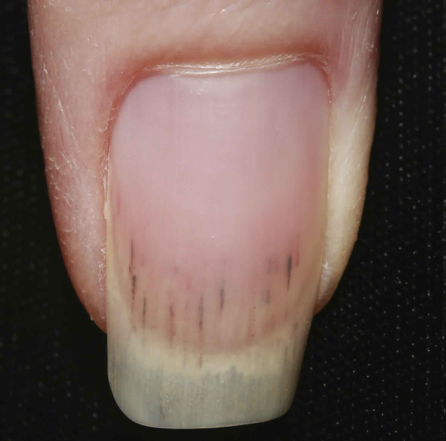
Clinical Context: A photograph of a patient with endocarditis, showing Osler nodes in conjunction with other signs such as Roth spots (retinal hemorrhages with white or pale centers) and splinter hemorrhages (linear hemorrhages under the nails).
Evolution of Osler Nodes: A series of images depicting the evolution of an Osler node over time, from its initial appearance as a small, painful nodule to its resolution with appropriate antibiotic treatment.
Differential Diagnoses: Images illustrating differential diagnoses for Osler nodes, such as traumatic lesions, vasculitis, or other types of infectious or autoimmune conditions that can cause similar skin manifestations.
Osler Node in Atypical Locations: Pictures of Osler nodes in less common locations, such as the toes or fingers, highlighting the variability in their presentation.
Osler Node with Other Signs of Endocarditis: An image showing an Osler node in conjunction with other peripheral signs of endocarditis, such as clubbing of the fingers or a new heart murmur, emphasizing the importance of a comprehensive physical examination.
Resolution Post-Treatment: An image of an Osler node after successful treatment of endocarditis, demonstrating resolution of the lesion and highlighting the importance of prompt and appropriate antibiotic therapy.
Imaging Studies: Images from imaging studies such as echocardiography or MRI, which can be used to visualize the heart valves and confirm the diagnosis of endocarditis in patients with Osler nodes.
Microbiological Confirmation: A photograph of microbiological cultures or PCR results confirming the presence of a pathogen known to cause endocarditis, such as Streptococcus viridans or Staphylococcus aureus, in a patient with Osler nodes.
Conclusion
Osler nodes are a critical diagnostic sign of endocarditis, a serious and potentially life-threatening condition. Understanding their clinical appearance, significance, and differentiation from other skin lesions is essential for medical professionals. The use of images, as described, can enhance medical clarity and facilitate earlier diagnosis and treatment, improving patient outcomes.
FAQ Section
What are Osler nodes, and why are they important in medicine?
+Osler nodes are painful, indurated skin lesions that appear on the extremities and are a diagnostic sign of infective endocarditis, a serious infection of the heart valves.
How do Osler nodes differ from Janeway lesions?
+Osler nodes are painful and indurated, whereas Janeway lesions are non-tender and appear as erythematous or hemorrhagic macules on the palms and soles.
What is the significance of Osler nodes in the diagnosis of endocarditis?
+Osler nodes are one of the peripheral signs that, in conjunction with other clinical findings and diagnostic tests, can confirm the diagnosis of endocarditis, guiding appropriate treatment.
How are Osler nodes treated?
+Treatment of Osler nodes involves addressing the underlying infective endocarditis with appropriate antibiotic therapy. Resolution of the nodes typically follows successful treatment of the infection.
Can Osler nodes appear in atypical locations?
+While Osler nodes most commonly appear on the palms and soles, they can occasionally be found in other locations, such as the toes or fingers, though this is less common.
What is the role of imaging in the diagnosis of Osler nodes and endocarditis?
+Imaging studies, particularly echocardiography, play a crucial role in diagnosing endocarditis by visualizing the heart valves and any vegetations that may be present, while also assessing cardiac function.
